What it is
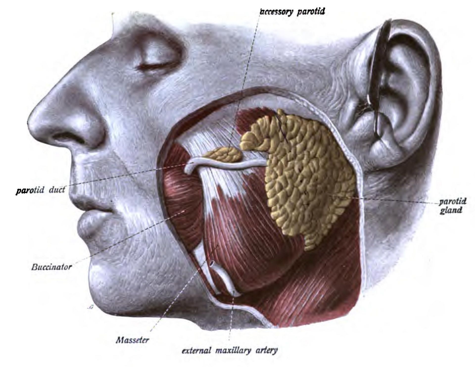
You have three pairs of major salivary glands: the parotid glands in front of the ears, the submandibular glands under the jaw, and the sublingual glands under the tongue. Each drains saliva into the mouth through a fine duct.
Minerals in saliva can form a stone in the duct, rather like a kidney stone. A duct can also narrow after repeated inflammation. Either blocks the flow. Most stones form in the submandibular gland, whose saliva is thicker and whose duct runs upwards to the mouth.
Symptoms
The typical sign is swelling and an ache that begin with eating, when saliva flow increases, and go down over an hour or two. Some people notice a salty or gritty taste, or can feel a hard lump under the tongue.
If saliva stays trapped, the gland can become infected. It is then constantly swollen, tender and red, pus may come from the duct into the mouth, and you may have a fever.
When to see an ENT surgeon
See a doctor if the swelling keeps returning, does not settle, or the gland becomes infected. A lump in a salivary gland that is always there and does not change with meals is not typical of a stone and needs examination, as does any weakness of the face.
Go to an emergency unit if swelling spreads into the floor of the mouth or the neck, or you have difficulty swallowing or breathing.
How it is treated
Small stones sometimes pass on their own with plenty of fluids, sour sweets to increase saliva, and gentle massage of the gland towards the mouth. Infection is treated with antibiotics. An ultrasound is usually the first scan.
When the blockage remains, sialendoscopy allows a scope about a millimetre wide to be passed into the duct to remove stones and widen narrowings, keeping the gland. Dr Kana has published a ten-year review of the technique. Removing the gland is kept for cases where this does not work.
Common questions
Is a salivary stone dangerous?
Not usually, but repeated blockage can lead to infection and gradual damage to the gland, which is why persistent symptoms are worth treating.
Which scan will I need?
Ultrasound is usually the first test. A CT scan is sometimes used to show the size and position of a stone.
Will I lose the gland?
Most people treated with sialendoscopy keep the gland. Removal is considered when the scope cannot clear the blockage or infections keep returning.
This page is general information, not medical advice for your situation.